Context
Since the beginning of this century and thanks to the development of advanced imaging technologies and probes, we can observe the molecular dynamics and interactions in live cells at both the microscopic and the nanoscopic scales. Dynamic imaging in microscopy is then exploited as an investigation tool to understand the function of particular biomolecules of interest. However, the mechanisms of life are commonly driven by multimolecular interactions and most of these interactions take place within ``molecular machines'' resulting of the assembly of multiple macromolecular species. Such ``machines'' are submitted to a continuous ``building-destruction'' process. They insure key steps of the biology of the cell and their dysfunctioning necessarily induces pathological situations. Molecular structures of the cytoskeleton such as the mitotic spindle, the cell signaling complexes, the nuclear envelop, the structures involved in intracellular transport, those responsible for adhesion or for cell morphogenesis and motility, all illustrate the notion of dynamic molecular assembly. Biological gatherings exist at different scales, (molecular, subcellular and cellular, at the level of tissue and beyond) while being often maintained outside a thermodynamic equilibrium. In all cases, they constitute sub-systems or integrated modules whose activities are themselves controlled by signalization and/or regulation via a large number of molecular networks. Then, a dynamic, quantitative and integrated description of molecular interactions within macromolecular complexes at the cellular level appears today essential for the global understanding of live mechanisms. This global approach also related to the emergence of systemic biology, which can be defined as part of the natural extension of the genomic era, will be necessary to identify the processes resulting in pathological situations (cancer, degenerative diseases), as well as to validate future therapeutic agents.
The present project will focus the Rab protein family involved in the membrane transport. This family of small GTPases (more than 60 in human), plays multiple roles via their association to internal membranes. Each member of this family exists under different dynamic states in the cell: they exchange continuously between the cytosol and the target membrane of one specific organelle, the donor compartment. They are associated to transport intermediates which are routed along the cytoskeleton networks. Finally, they may associate, most probably transitory, to their membrane of destination, the acceptor compartment. Their functions are regulated by a cycle of GTP hydrolysis as well as by their molecular interaction with a large number of partners, in the context of multiprotein complexes. The analysis of the assembly of these complexes, the dynamics of their assembly and the role of Rab proteins inside those complexes are fundamental for the understanding of the molecular mechanisms responsible for membrane transport and for the maintenance of the cell integrity. Moreover, the interconnectivity and the spatial organization of such protein systems support the basic cellular functions and must be considered in the context of the global architecture of the biological unit: the cell. Then, cellular functions must be thought as an integration of molecular activities in space and time. This constitutes one of the challenges of the modern biology and it becomes imperative to develop new approaches and to reinforce the technologies that are necessary for the measurement of the dynamics of those biochemical processes in ``intact'' cells. Today, it is one of the main concerns at the Institut Curie and more particularly in the team ``Mécanismes Moléculaires du Transport Intracellulaire'' at the UMR 144-CNRS.
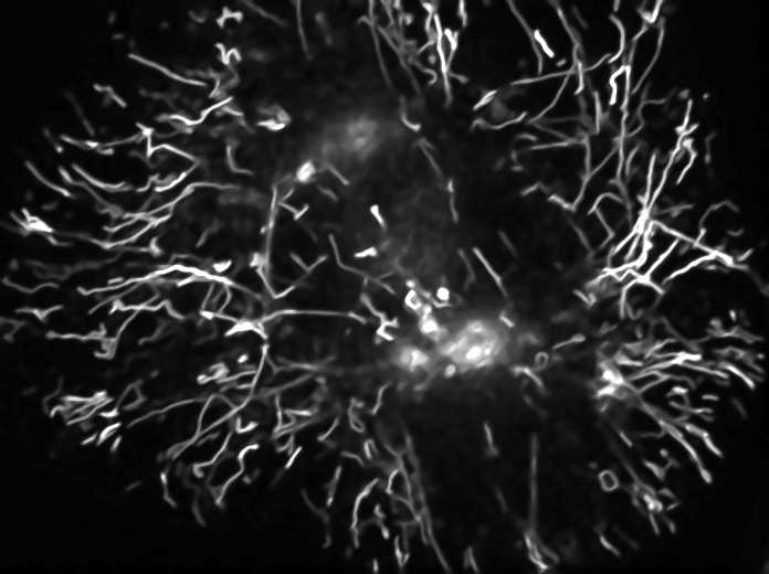
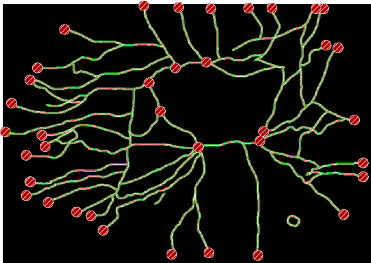
This last decade, advances in XFP-tagging and time-lapse microscopy have revolutionized intra-cellular and molecular biology and have opened new avenues to study the dynamics and the function of multiple proteins. The nD images are constituted by a large number of dimensions (two or three spatial dimensions and one temporal dimension for each spectral channel considered and corresponding often to only one particular biomolecular species). Dynamic microscopy is also characterized by the nature of the observable objects (cells, organelles, single molecules,...), eventually by the large number of small size and mobile elements (chromosomes, vesicles, ...), by the complexity of the dynamic processes involving many entities or group of entities sometimes interacting, by particular phenomena of coalescence often linked to image resolution problems, finally by the association, dissociation, recomposition or constitution of those entities (such as membrane fusion and budding). Thus, the corpus of data to be considered for a comparative analysis of an experiment constituted by multiple image series acquisitions is massive (up to few Giga-bytes per hour). Consequently, this corpus becomes particularly difficult to qualitatively and quantitatively interpret manually; most commercial image analysis tools for automatically extracting information are rather limited and/or require a large amount of user interactions. Moreover, data-sets that could be exploitable are insufficiently standardized, making them difficult to compare. Therefore, it becomes absolutely necessary in a wide number of modern biology and biomedical fields to facilitate and rationalize the production of those nD data, to improve post acquisition treatments (e.g. image processing) which are limiting factors in front of the data flux, and to favor the organization and the interpretation of the information associated to this data corpus.
Image sequence analysis in video-microscopy for life sciences has not been investigated by the past by computer vision groups, but now has gained importance since molecular biology is presently having a profound impact on the way research is being conducted in medicine. One can see the constitution of teams concerned by the acquisition, treatment and analysis of dynamic image data. Moreover, networks at the European level [EAMnet for European Advanced Microscopy Network (since 2003) ; ELMI for European Light Microscopy Initiative (since 2001) ; French/German Initiative for Biophotonics (since 2005)] but also at the national level [GDR 2588 ``Microscopie du Vivant'' (since 2002) ; RT ``Microscopie Fonctionnelle et Multidimensionnelle'' (since 2004)] attempt to gather the research efforts made in this multidisciplinary context. Most of the partners of the present proposal are active members of these networks. This motivates our present research effort which is to develop novel approaches based on recent techniques in computer vision and signal processing but also in modeling and mathematical biology to extract quantitative measurements from nD data related to intracellular dynamics in biology. The current techniques (particle filtering-based tracking methods, ...), are not able to process complex data corresponding to interactions between several dozens of moving objects with variable velocities. In addition, analytical models of movements are poorly adapted for such complex situations. For those reasons, new developments and other parsimonious models should be envisaged. They also rely on the adaptation of acquisition tools, the increase of post acquisition treatment robustness and efficiency aiming at improving the image resolution and contrast.