Demo bi-transparency - Fluoroscopic image sequence 19
(back)
Description
We observe on this example the heart (the grey mass present in almost all of the image), superimposed to bright lung
tissues. The (deep dark) diaphragm is visible at the bottom of the image, and diagonal ribs also appear.
Under this angulation of radiation, the heart appears static, so that it can be merged with the ribs in a first layer.
The second layer, animated by the breathing, includes the lungs and the diaphragm.
Let us point out how the motion-based definition of layers is adapted to our problem, in contrast to anatomical definitions.
In this sequence, the clinician moves the whole imagery system during the acquisition in order to center it on the region
of interest. During this operation (the panning), the scene undergoes a global translation.
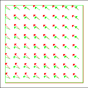
The estimated displacements are directed toward the past to respect the notations ot the Transparent Motion Constraint.
As a result they are in the opposite direction compared to the usual represented velocity direction.
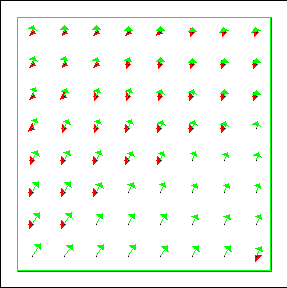
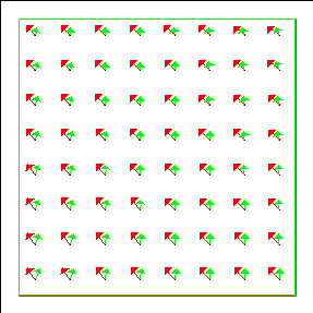
Estimation result at four interesting time instants
The first presented image refers to a time instant before the panning, where the motions of the two layers are
correctly estimated.
The three other images correspond to successive time instants during the panning. We can note
that the global motion is correctly handled, and that the estimated motions models are satisfactory. This example shows
also that our transparent motion estimation method is able to correctly compute relatively large motions.
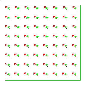
Whole processed sequence
We also present the overall processed sequence. It shows the estimated affine motion fields, along with the corresponding
frames.
The results are satisfactory for about 70% of time instants. Note that when both layers are static, the estimation
method still estimates two affine motion fields. This results in a correctly static motion field and a second
incoherent estimated motion that corresponds to the noise.
|
|